Neurobiologist Rodal wins New Innovator award
Interplay between neuronal firing and membrane traffic to be investigated
 Photos/Charles A. Radin
Chances are you didn’t spend much time this morning getting your shoes on or thinking about which route you would take to work. But for people suffering with debilitating diseases such as ALS (Lou Gehrig’s disease) or Alzheimer’s, such simple functions -- and even activities like breathing and eating -- can be a struggle.
They are the people that Avital Rodal, assistant professor of biology, hopes to help through research that has just been funded by the National Institutes of Health. She hopes her work, which asks how neuronal firing affects transport of materials within neurons, will someday provide not only answers, but lead to solutions.
Rodal is one of the 51 NIH Director’s New Innovator award recipients for 2012. The awards are given to researchers to pursue visionary science that exhibits the potential to transform scientific fields and speed the translation of research into improved health. The High Risk High Reward program is supported by the National Institutes of Health Common Fund. Rodal’s lab will receive $300,000 per year for five years, plus additional research support costs for the university.
 |
|
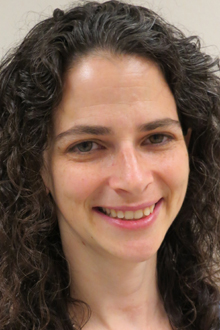
| Avital Rodal |
Rodal’s lab works on how packets containing growth and survival signals are transported inside cells in membrane compartments. That is crucial for neurons, she says, as nerve cells can reach from the spinal cord all the way down to the toes.
“If you’re a blue whale, that can be over 20 meters,” says Rodal. “ There is a whole highway system that packages these growth signals into little taxis that can then move along tracks that can bring them to another part of the neuron.”
The body has multiple neurons that are in a relay from your brain to your spinal cord, then from your spinal cord to, say, your finger. Says Rodal, your finger is secreting growth factors, which stick to receptors on the membrane. Those receptors communicate back to the neuron by pinching in, so the membrane actually makes a little vesicle, which used to be the edge of the cell. Once pinched in, this vesicle contains and growth factors
In studying how growth factors are internalized into the cell and how they move through the neuron, Rodal’s lab has developed new microscopy techniques that give them the ability to observe the process in real time in fruit fly neurons.
Her hypothesis is that electrical activity in neurons lead to a change in how growth factor receptors are moving back and communicating to the neuron, telling it whether it should grow, survive, or become stronger or weaker.
“What I wanted to do in this project was to draw the connection between firing of the neuron and traffic of these receptors,” says Rodal. “I’m in a good position to do that is because I have this great model system where I can make neurons fire the way I want them to, and I can look at the receptors trafficking.”
Rodal wants to know: does a neuron that fires a lot shunt its growth factors faster, or is it something completely different? The other part of her project involves purifying the molecular machines that drive these different methods of transportation to see how they are different between neurons that are firing frequently versus infrequently. This work will pinpoint strategies that healthy neurons use to alter growth signaling, giving researchers new ideas for therapeutics to correct growth signaling defects in neurodegenerative disease patients.
“In Lou Gehrig’s disease, motor neurons that innervate your finger die, in part because they lack these growth factors,” says Rodal. “In neurodegenerative diseases, the molecular machines that drive membrane traffic go wrong. We’d like to be able to turn growth factor signaling back into a healthy state, and to do that, we need to know what’s happening to each one of these machines that moves things from one compartment to another.”
Categories: Research, Science and Technology