Rodal explores ‘eye candy’ with Pew grant
Biology professor studies neuronal systems and neurodegenerative diseases
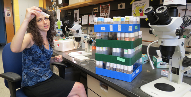 Photos/Charles A. Radin
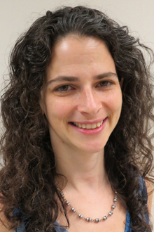
Avital Rodal
Rodal, who was recently named a Pew Scholar in Biomedical Sciences, studies how neurons set up elaborate structures tailored to send and receive electrical signals over distances and through complex networks.
“I just love being in the lab and having an idea and testing it and making discoveries,” she says. “I’ve just always thought biology was cool, as far back as high school. But I don’t want to just learn about it; I want to do it. I can’t believe people pay me to do this.”
“It’s a tough job and it’s very competitive because resources are limited and you have to fight really hard for it,” she adds.
 “The way we maintain academic rigor is by constantly criticizing each other, so it’s not a feel-good job.”
Rodal, a faculty member at Brandeis since 2010, is winning the fight. The Pew Scholar designation brings with it $240,000 to continue her neuroscience research for the next four years. Last year, she won the NIH New Innovator Award and the March of Dimes Basil O'Connor Starter Scholar Research award.
An Ottawa, Canada, native, Rodal came to the Boston area to pursue her undergraduate degree at MIT, then headed to the West Coast in 1997 to earn a PhD at Berkeley.
There she studied the inner structure (or cytoskeleton) of cells with David Drubin, and decided to, “take what I learned in my PhD and apply it to neuronal systems” – which she did when she returned to MIT to do a post-doc in Troy Littleton’s lab. When her then-boyfriend (now husband), Biology Professor Bruce Goode accepted a faculty position at Brandeis, she came as a visiting scientist and helped him set up his lab and made many connections.
“That’s when I got to know how awesome Brandeis was,” she says with a broad smile. “All of these people who were doing this awesome science, but together in a collaborative and congenial way, which is not the case in a lot of institutions. In many other places, each lab is kind of its own kingdom and there isn’t a lot of interaction between them. It’s very isolated and isolating.”
Rodal was happy to return to the collaborative environment in 2010 when she finished her post-doc because “it’s very multidisciplinary. The barriers are nonexistent.”
A case in point, she says, is how six faculty members from different departments jointly applied for and won a grant to purchase a microscope that Rodal calls a “unique, completely custom system we use to actually watch these intracellular trafficking events occurring, so you can really see them happening in a living animal and in living tissue.”
As she talks about it, experts outside her office are installing a new component to the microscope, which is up and running, she says. She quickly turns to her computer, asks, “Want to see what it does?” and gives a show-and-tell of videos it and other instruments obtained. “It’s great eye candy,” she says.
Specifically, Rodal’s research focuses on growth factor signals. For example, the tissue in your finger secretes growth factors that are taken up by the neuron that activates movement in your fingers. These growth factors are transported in membrane-bound packets up your arm and to your spinal cord to promote neuronal survival.
Much of the work in the lab explores processes associated with neurodegenerative diseases. If those signals are depleted because your finger is no longer secreting the growth factors or because something’s wrong with the transport, that neuron dies and you may get a neurodegenerative disease like ALS or Alzheimer’s. Her lab has emulated, observed and manipulated these processes in flies.
With the Pew funding, her lab will mix and match proteins that act like scissors and pinchers on membrane-bound growth factor packets, to see how they work together. Because these membrane-bound packets of proteins are so tiny – hundreds of nanometers across – they are difficult to study with a conventional microscope. But Rodal proposed using new technology that allows researchers to “push the resolution of what you can see in a light microscope.”
Categories: Research, Science and Technology