A new dimension in understanding cilia
Researchers use 3-D imaging and modern electron microscopy to see cellular structures in high resolution
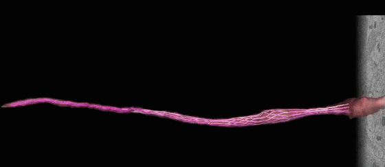
Reconstructed 3-D cilia in sensory neurons
You’ve never seen cilia like these before.
Brandeis University researchers have developed a new model to study these tiny but vital cellular structures with more clarity and detail than ever before, providing a clearer picture on how cilia are shaped, structured and how they interact with their environment.
The team, lead by professors of biology Daniela Nicastro and Piali Sengupta, published their findings in the journal eLife on March 25.
Cilia, antenna-like appendages of cells, come in many shapes and sizes, each fulfilling a different purpose. Some help propel cells or fluids along tissues, others have sensory functions, gathering information about the animal’s environment. Humans see, hear and smell using cilia — human olfactory neurons have a tuft of about six to 17 cilia that house smell-signaling proteins. Without effective cilia, cells and whole organ systems can malfunction, degenerate and die, leading to disorders such as anosmia (loss of smell), blindness or Polycystic Kidney Disease.
 |
| Reconstructed 3-D cilia in sensory neurons |
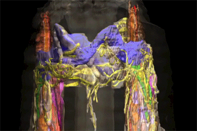
Nicastro and Sengupta, with postdoctoral fellows David Doroquez and Cristina Berciu, studied cilia in the sensory neurons in the noses of roundworms. To do so, the team needed to quickly preserve the worm without changing its cellular structure and take very thin slices off its nose. They employed a state-of-the-art technique that involved freezing the worms in a matter of milliseconds at a pressure almost twice as high as the deepest part of the world’s oceans. The team then slowly replaced the frozen water molecules with a preservative solution and thinly sliced the nose — cutting each slice about 1,400 times thinner than a sheet of paper.
They used transmission electron microscopy and electron tomography to acquire images of the ultra-thin sections and then fed these images into a computer to combine them into 3D reconstructions and models. More than 120,000 images were recorded for this study.
The results are mesmerizing — beautifully detailed 3-D models of various types of cilia extending from 50 neurons. Some cilia are simple rods, or two branches splitting from a shared dendritic trunk; others are more complex, shaped like leaves, vines, Weeping Willows or tree arbors. The high resolution images allowed for the discovery of dendritic branches that are 40 times thinner than an E. coli bacterium.
Understanding the spectrum of cilium shape and cellular structures can help scientists explore how these contribute to specific sensory functions and which genes control those unique architectures.
The research was funded in part by the National Institutes of Health and the National Science Foundation.
The full paper with movies is available on eLife.
Categories: Research, Science and Technology