Diomedes Logothetis, PhD
Professor
Department of Pharmaceutical Sciences
Northeastern University
(February 25, 2020)
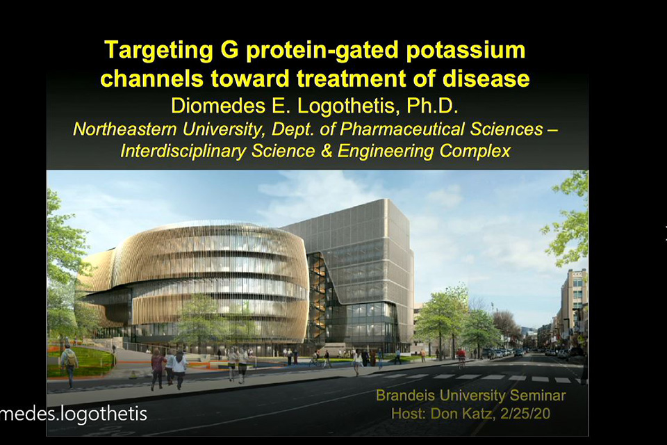
Targeting G protein-gated potassium channels toward treatment of disease
An issue with many medications is that they do not target only one area of the body. Antihistamines, for example, can block an allergic response, but they also block histamine receptors in the brain, leading to sleepiness. This can, as Dr. Logothetis explained, have more serious consequences. Treatments for atrial fibrillation (an abnormal heart rhythm) often interfere with normal heart rate variability. Treatments for addiction and anxiety can also have unwanted effects on the heart. Dr. Logothetis discussed his research on finding ways to target effects of medications to only where they are needed.
 Ion channels are proteins that span the plasma membrane and provide a hydrophilic path for ions to pass through from the inside to the outside of the cell and vice versa. These ion movements control the excitability of cardiac or neuronal cells. Certain potassium (K+) channels expressed in atrial and pacemaking cells of the heart can be activated by Acetylcholine (ACh) that is released by the vagus nerve to slow down heart rate. These KACh channels are responsible for allowing the heart to adapt rapidly to varying cardiac output demands by controlling heart rate. Thus, heart rate variability (HRV) serves as an index of good cardiac health. Yet, overactivity of KACh channels, as occurs under oxidative stress conditions during aging, gives rise to the most prevalent cardiac arrhythmia, atrial fibrillation (AFib), encountered in 9% of the senior citizen population. A life-threatening complication of AFib is blood clot formation in the atria and high risk for stroke, a condition requiring patients to chronically take blood thinners, which have their own complications of risk for hemorrhage. On the other hand, neurotransmitters like opioids, dopamine or GABA that activate these channels that are also expressed in the brain are involved in countering pain, addiction and anxiety.
Drug discovery efforts to produce inhibitors that correct the overactivity seen in AFib but do not compromise HRV have not succeeded. Similarly, it has not been possible to make specific activators of these K+ channels in the brain, as opposed to the heart. I have been studying KACh channels for the past 35 years. We have reasoned in my lab that understanding the molecular details of activation of these channels could allow us to eventually design drugs that could dial down their overactivity without inhibiting them completely in heart or to specifically activate brain over cardiac channels. ACh activates the channels through the signaling proteins referred to as GTP-binding (G) proteins that couple muscarinic receptors to KACh. In fact, we found that it was the Gbg dimer that interacts directly with KACh to activate it. Furthermore, we discovered that intracellular Na+ ions could also activate KACh independently of G proteins but could also synergize with Gbg to cause maximum activation. The channels expressed in the heart utilize different subunits (GIRK1/GIRK4) from those expressed in neurons (GIRK1/GIRK2) and possess a long permeation pathway with two gates, one at the inner leaflet of the plasma membrane and the other at the cytosol. We discovered that a signaling phospholipid, phosphatidylinositol-bisphosphate (or PIP2), was a key component of the plasma membrane controlling channel opening and Gbg or Na+ somehow changed the protein conformation to modulate its strength of interaction with PIP2 and control gating.
In the past decade high-resolution crystal structures and models of GIRK channels with Gbg, Na+ and PIP2 have been determined. We have developed computational dynamic models that in hundreds of nanosecond simulations reproduce gating of GIRK channels by Na+, Gbg, and the two together, in the presence but not the absence of PIP2. The models have predicted that the weaker activation of Na+ ions is due to activation of the cytosolic gate, the stronger activation by Gbg is due to activation of the inner membrane gate, while the synergism between the two activators comes from simultaneous activation of both gates. High fidelity homology models of the brain (GIRK1/GIRK2) or the cardiac (GIRK1/GIRK4) channels have also produced in molecular dynamics simulations the proper physiological behavior in response to the channel gating molecules (Na+, Gbg and PIP2). Having developed these powerful computational models we have been able to simulate the effects of small molecule drugs and through structure-based drug design approaches to open specifically the brain but not the heart channels. Such small molecule activators have been effective in rat fear extinction models of post-traumatic stress disorder. Simple chemical modifications of these drugs switch their action from activators to inhibitors and their specificity from the brain to the cardiac channels. We anticipate that with our structure-based drug design approach we will succeed in producing the needed partial inhibitor of KACh that could treat aging-dependent AFib.
