2014 Research Highlights
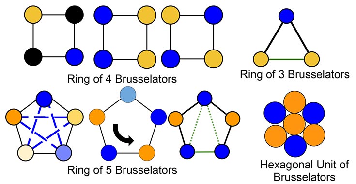
Upper Left: Ring of 4 Brusselators. Upper Right: Ring of 3 Brusselators. Lower Left: Ring of 5 Brusselators. Lower Right: Hexagonal Unit of Brusselators.
Theoretical analysis of emergence of patterns in an initially homogenous ring of chemical oscillators exhibits a multitude of patterns. The chemical reactions are modeled by the Brusselator, which is one of the simplest autocatalytic reactions, in the regime of high nonlinearity. They are coupled diffusively. The emergent heterogeneity is characterized by the oscillator phase (color in diagrams). A black circle denotes an oscillator that has stopped oscillating.
NSF MRSEC 0820492
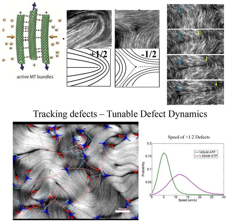
Upper left diagram: active MT bundles. Upper middle: a pair of diagrams labeled "Characteristic Defects of a Nematic Liquid Crystal." Upper Right: "Unbinding of a pair of defects." Lower left image: "Tracking defects -- tunable defect dynamics. Lower right: Speed of + 1/2 Defects.
While conventional materials are assembled from inanimate building blocks, we are exploring the behavior of soft materials in which the constituent components consume energy and spontaneously coordinate their microscopic behavior and form novel materials such as active gels, crawling emulsion droplets and living liquid crystals.
In active liquid crystals, defects proliferate to a dynamical steady state through unconventional creation and annihilation processes. Pairs of defects stream across the nematic liquid crystal exhibiting tunable dynamics not readily observed in equilibrium liquid crystals.
Brandeis MRSEC (0820492)
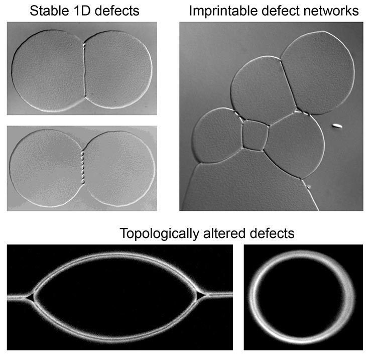
We have discovered two novel frustrated coalescence pathways in colloidal membranes that result in the formation of stable liquid crystalline defects. This frustration propagates up several hierarchical levels, beginning with the molecular chirality inherent to the constituent virus particles. We observe the structure of these defects using optical microscopy, measure the energetic cost of their formation, and imprint arbitrary arrays of these defects through optical tweezer-driven membrane self-coalescence.
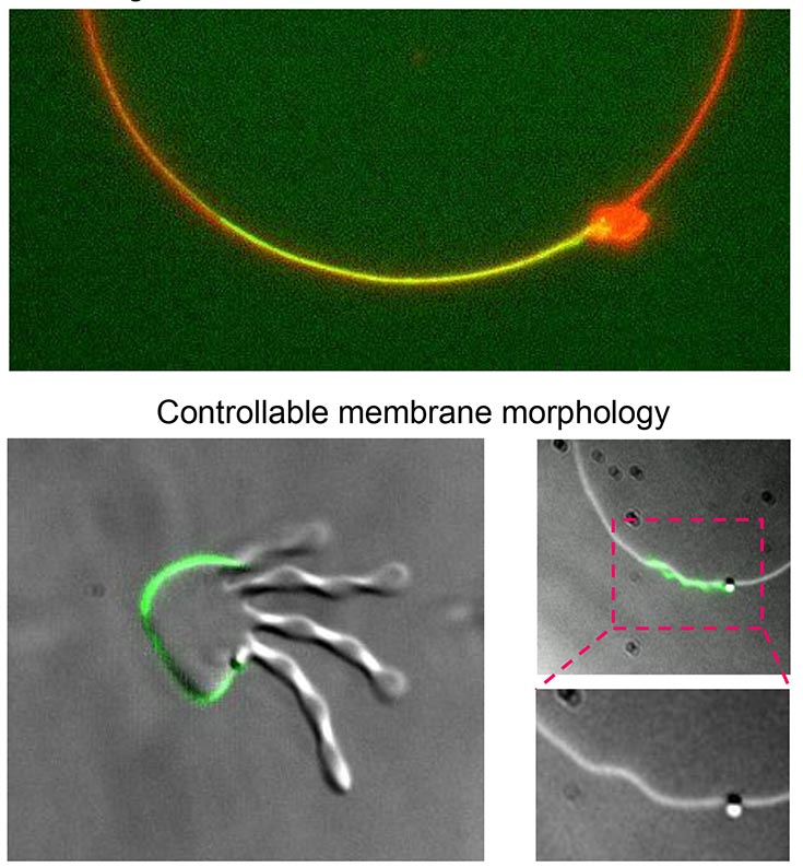
Top image: Novel geometrical method of interfacial association. Bottom images: Controllable membrane morphology .Three grayscale images of organic shapes with red and green markings on them.
The chemically heterogenous nature of typical surfactant molecules drives them to the interface between various immiscible fluids. We have discovered a new class of edge-active agents, homogenous filamentous polymers which preferentially adsorb onto the unique 2D interface of colloidal membranes due to geometrical considerations only. We use these geometrical edgeactants to predictably modify the bending rigidity of colloidal membranes by more than an order of magnitude, and to control the conformation and morphological behavior of the membranes.
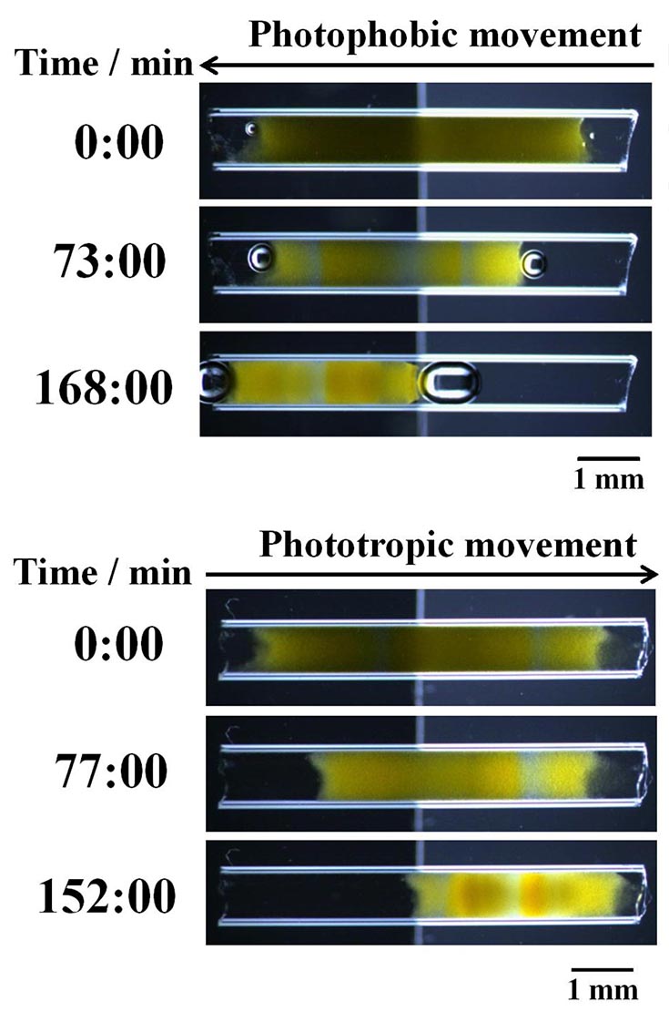
Upper image: Photophobic movement. Lower image: Phototropic movement.
Gels containing the components of the photosensitive oscillatory Belousov-Zhabotinsky reaction have been predicted to move toward darker regions when illuminated by a patterned light source. We have demonstrated experimentally that, with appropriately chosen patterns of light intensity, these gels can move either toward or away from brighter regions, mimicking the behavior of phototropic and photophobic organisms.
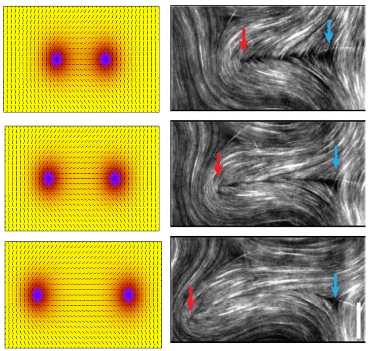
We are developing a dynamical theory of an overdamped active nematic. This will enable us to identify universal mechanisms for dynamical phenomena in diverse active nematic systems. The image on the right shows three sequential images from experimental system in which +½ and -½ defects are created through a bending stability and subsequently separate. The image on the left shows similar defect behaviors observed in the numerical solution of the non-equilibrium dynamical theory.
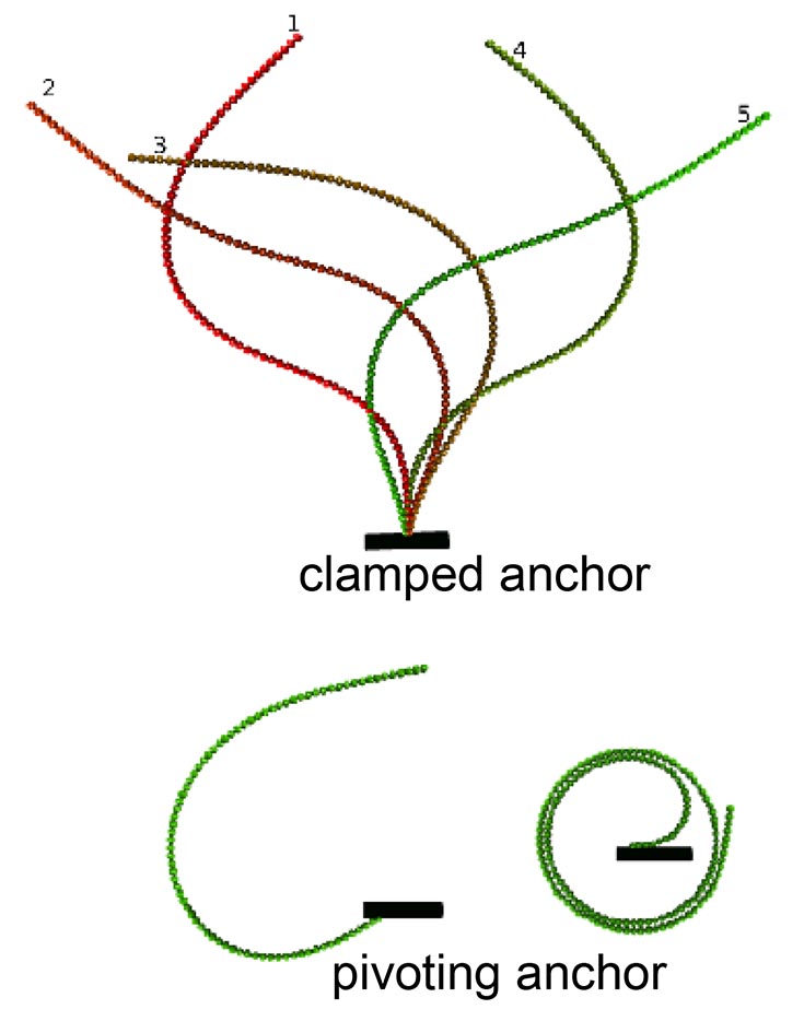
Filament configurations at different times are shown in different colors. Upper diagram: Clamped anchor. Lower diagram: pivoting anchor.
The planar dynamics of a semi-flexible filament anchored at one end and comprised of connected, self-propelled, spheres were predicted using Brownian dynamics simulations and continuum elastic theory theory. For certain parameter ranges the filament undergoes periodic motion. With a clamped anchor, the filament undergoes flagella-like beating (top right), while a pivoting end leads to a steadily rotating coiled conformation (bottom right).
Designing simple, experimentally feasible systems that mimic the periodic beating of eukaryotic cilia and flagella has important implications for controlling fluid flow at the microscale, as well as for understanding biological cilia and flagella.
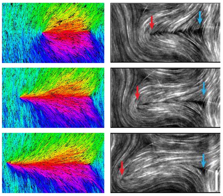
Simulations of a model for microtubule (MT)-based active nematics capture experimentally observed defect dynamics. The image on the right shows three sequential images from experimental system in which +½ and -½ defects are created through a bending stability and subsequently separate. The image on the left shows similar defect behaviors observed in dynamical simulations of a coarse grain model for the MTbased active nematics.
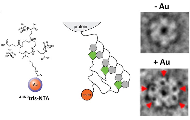
Structure 2014; 22(4): 628-635
Novel reagent consisting of a gold cluster or nanoparticle conjugated to a tris-NTA scaffold (called AuNP-tris-NTA, see figure). This reagent has been employed to localize protein(s) of interest in large multi-protein assemblies as shown in the negative-stain electron microscopic image where the gold nanoparticles sitespecifically aGached to the protein appear as dense black spots due to their significant scaGering of electrons as a consequence of the gold’s electron dense structure.
![]()
A National Science Foundation sponsored Material Research Science and Engineering Center
![]()