Image Competition
At the end of each year, Brandeis hosts an image competition for users of the shared facilities. Users submit their best art captured using facility microscopes and the winners are selected, printed and displayed in the facility. The image competition is a great intersection of art and science.
2024 Winners
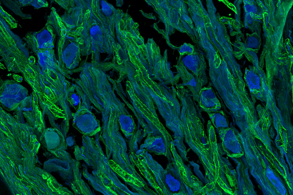
First Place: Acquired on the LSM880, this is a section of superior cervical ganglia stained for S100b (green) and Map2 (blue). The image shows clear glial envelopes around cells and wrapped around neuronal fibers running through the sample longitudinally and cross-sectionally.
Photo Credit: Ellie Greene, undergraduate student, Birren Lab
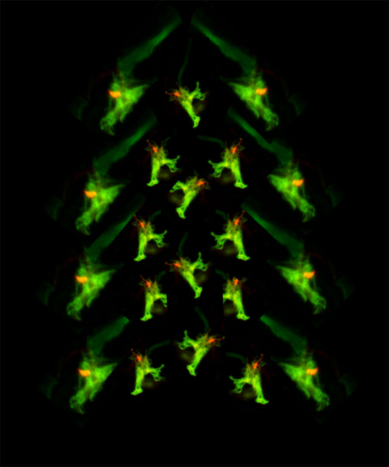
Second Place: Acquired on the LSM880 Airyscan, the images are of the sensory ending of the AFD neuron of C.elegans, with the Microvilli (green) and Cilium (red) arranged into a Christmas tree.
Photo Credit: Priya Dutta, postdoctoral researcher, Sengupta Lab
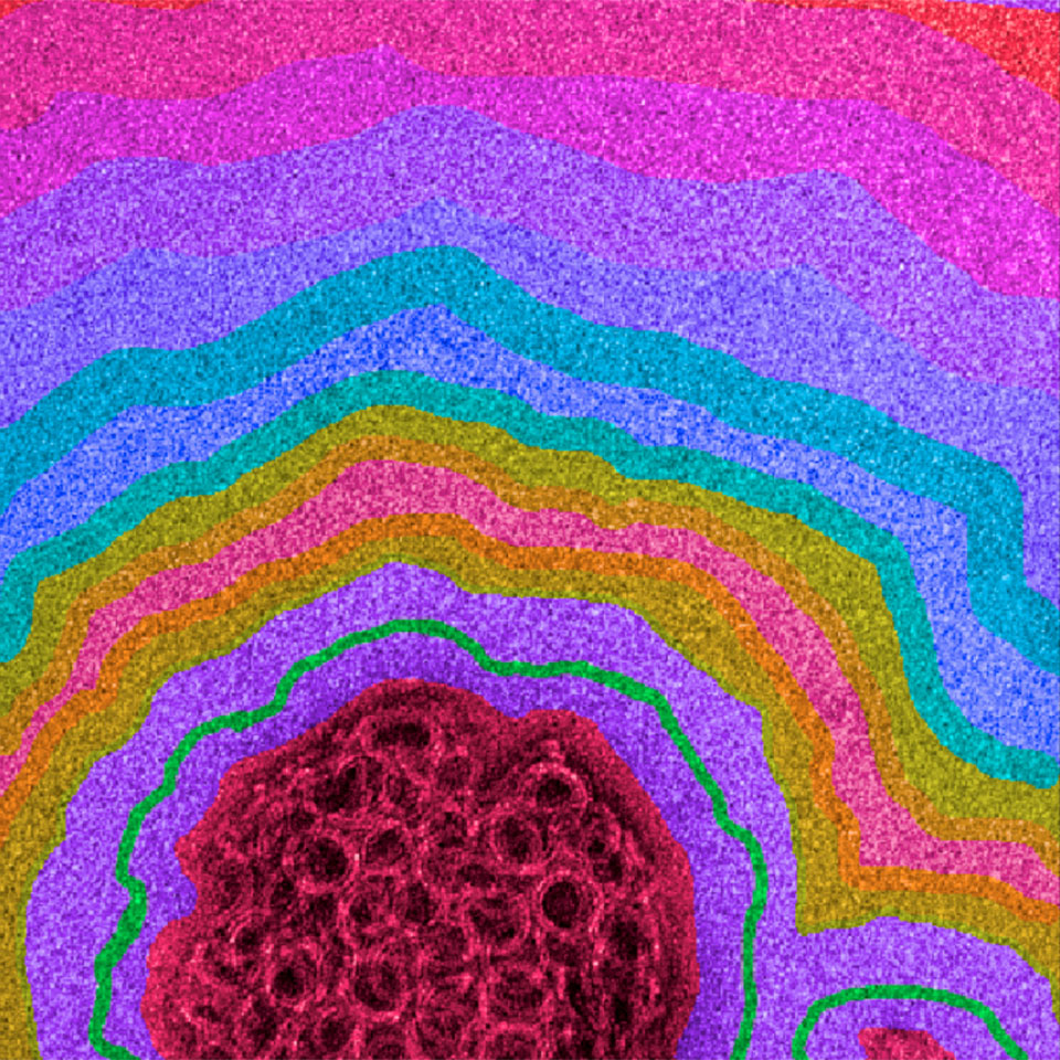
Third Place: This is a negative stain electron microscope image showcasing a pore-forming toxin (PFT) from Ralstonia Pickettii as it targets an artificial liposome. The original image was captured using a transmission electron microscope, with color enhancement applied in Affinity Design.
Photo Credit: Alexandra Hessberger, rotation student, Johnson Lab
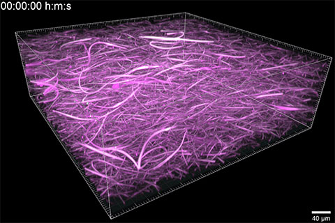
Best Video: Acquired on "Dagahra," the Nikon AX-R, these are asters with core-cortex structure self-assembled from a 3-D turbulent isotropic cytoskeletal.
Photo Credit: Quang Tran, Postdoc, Fraden Lab
2023 Winners
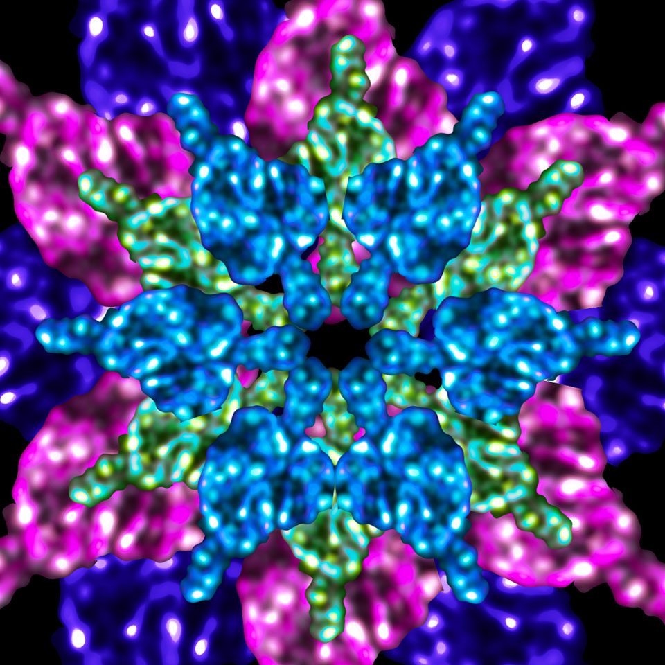
First Place: Acquired on the LSM880 Airyscan, these are boutons at the Drosophila neuromuscular junction turned into a kaleidoscope using Photoshop. Hidden in the kaleidoscope are the Drosophila proteins Shibire, Nervous Wreck and Bruchpilot.
Photo Credit: Anne Silveira, PhD student, Rodal Lab
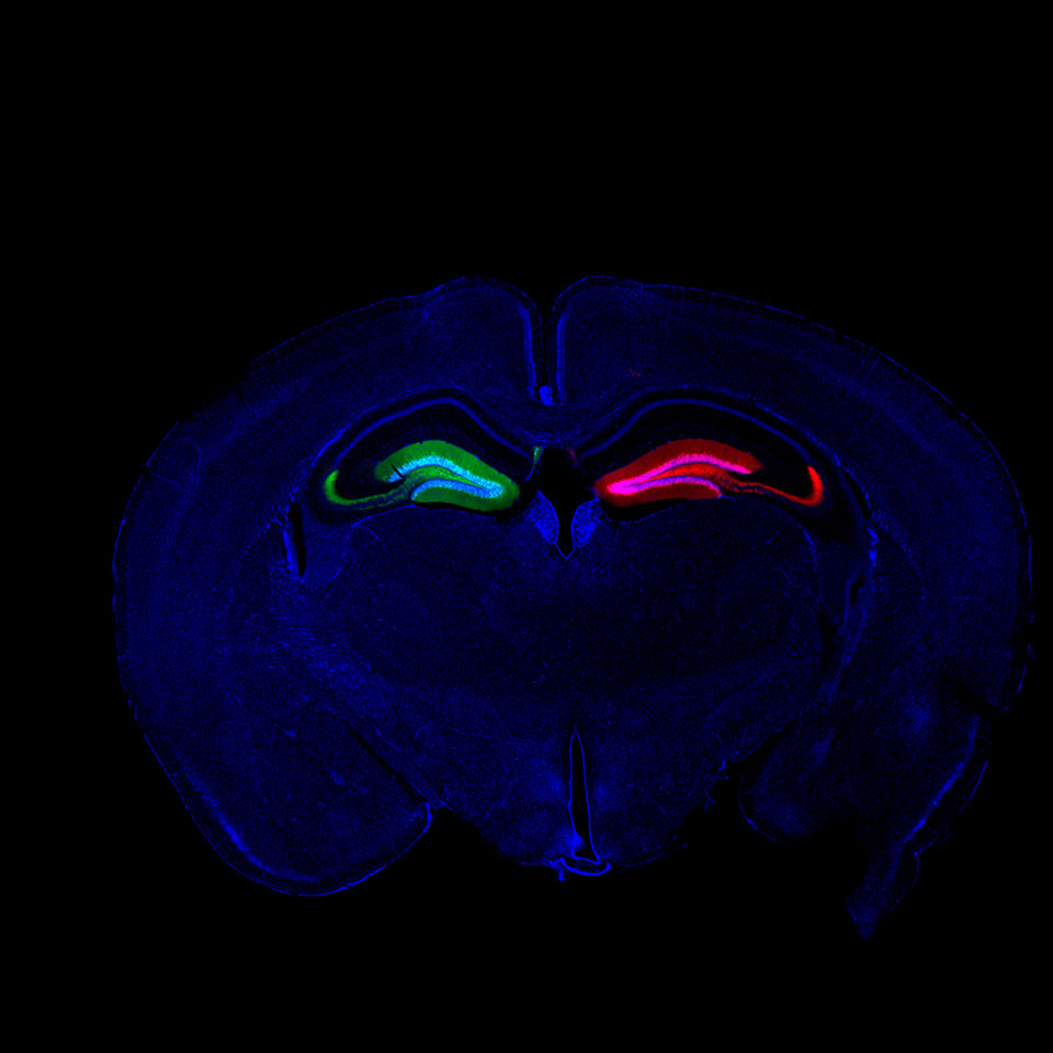
Second Place: Acquired on the Keyence, this is a coronal section of P30 mouse brain with unilateral Dentate Gyrus injections of Rfx3-Knockdown AAV (mCherry, red) and a non-targeting control AAV (EGFP, green).
Photo Credit: Ryan Kirk, PhD student, Nelson Lab
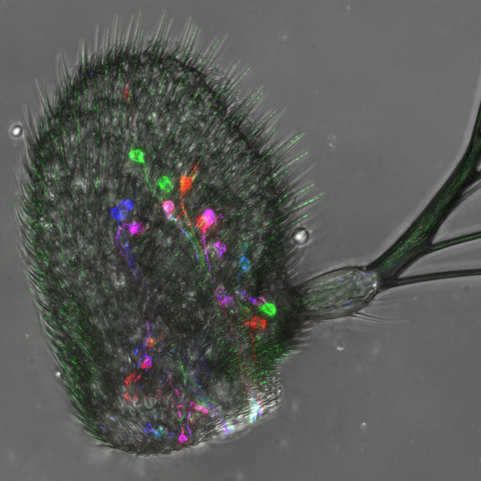
Third Place: Acquired on the Leica SP5, this image captures the expression of Rh50 (an ammonia transporter) in the antenna of Drosophila melanogaster. It uses MultiColor FlpOut (MCFO), a technique that can stochastic label cells. Green- FLAG tag, Red- V5 tag, blue- HA tag. In each of the Rh50 cell, 0 or 1 or 2 or 3 tags can be expressed by the combination of three colors. Cells can be labelled in 8 different ways. It is useful when cells have different subpopulations, and this technique can help trace where each population of cells project in the brain.
Photo Credit: Ruocong Tang, PhD student, Garrity Lab