Louise Mashal Gabbay Cellular Visualization and Electron Microscopy Center

"A 3.5A Cryo-TEM reconstruction of a bacterial pore-forming protein" Johnson Lab.
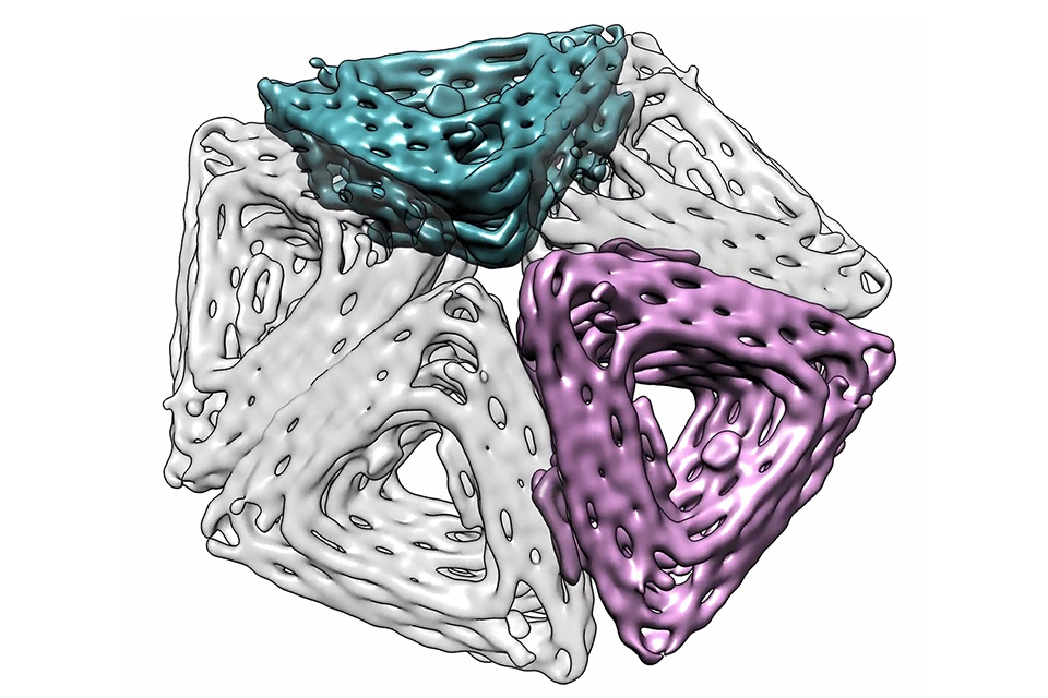
"A visualization using a cryo-TEM reconstruction of DNA origami subunits that can bind edge-to-edge, assembled into a pentamer, a step first towards assembling synthetic T1 capsids" Rogers and Fraden Labs.
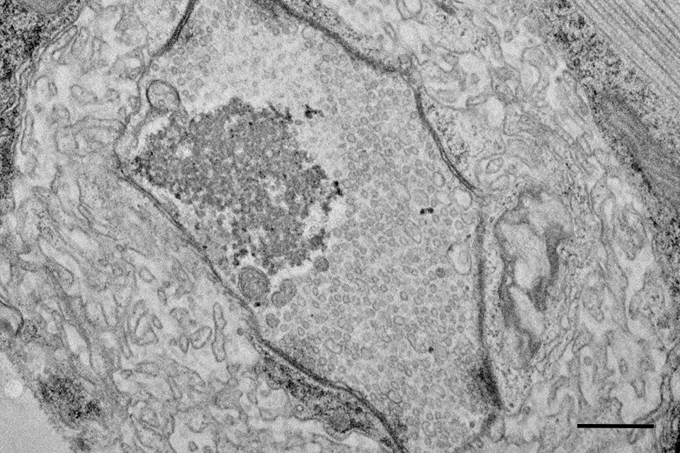
"Thin-section transmission electron microscopy of a synaptic bouton at the Drosophila larval neuromuscular junction. Scale bar 400nm" Rodal lab
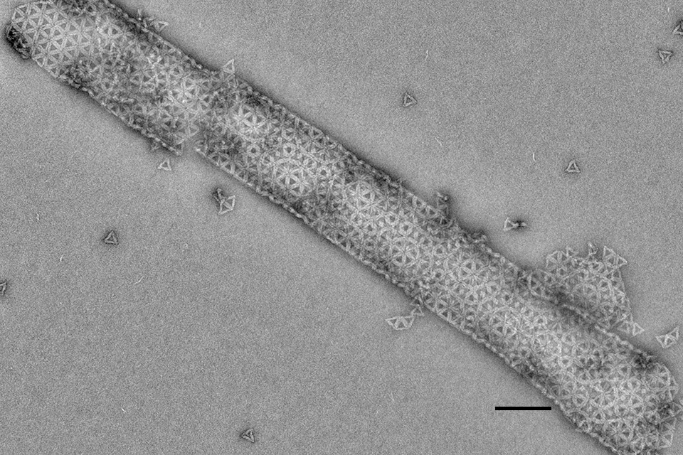
"Self assembled tubule of DNA origami triangles. Scale bar 200nm" Rogers and Fraden Labs
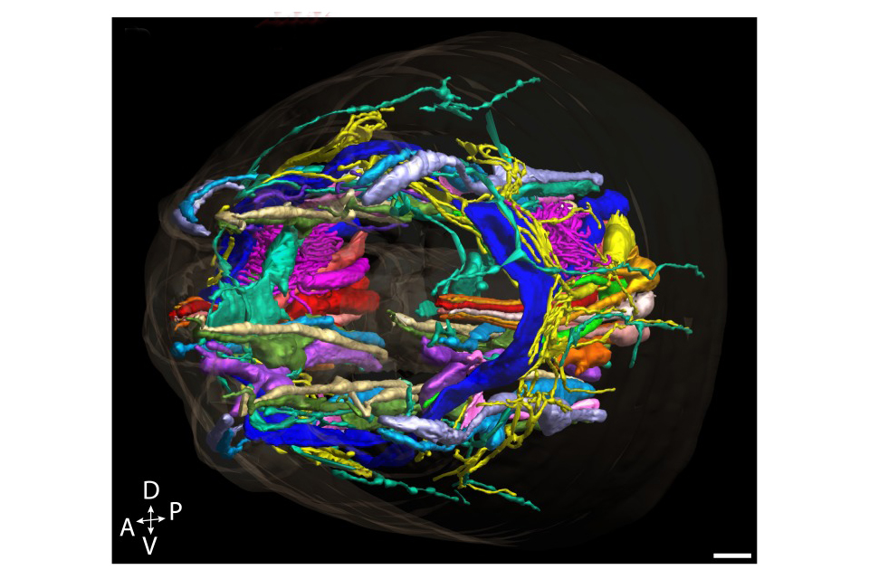
“A high-resolution morphological and ultrastructural map of anterior sensory cilia and glia in Caenorhabditis elegans.” Sengupta and Nicastro labs
The Brandeis Electron Microscopy Facility is located on the 4th floor of the Rosenstiel Basic Medical Sciences Research Center at Brandeis University in Waltham, Massachusetts, part of the Greater Boston area.
The facility is under the direction of Dr. Avital Rodal and is run by Dr. Berith Isaac.
Instruments, training, and assisted use are available for Brandeis faculty, staff, and lab members, as well as members of the broader scientific community, including other academic institutions and for-profit organizations.